By admin | Sep 07, 2016

The Vitreous Humor
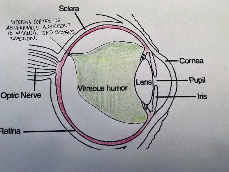
The vitreous humor is the gel-like substance which resides in your eye. The vitreous fills the space between the lens and the retina and is encapsulated within a thin shell called the vitreous cortex.
The vitreous cortex itself adheres to the retina. The vitreous acts to maintain the shape of the eye and acts as a shock absorber. For more information about the vitreous check out our previous blog post on Posterior Vitreous Detachments (PVD).
What is Vitreomacular Traction Syndrome?
Vitreomacular Traction Syndrome (VMT) can occur as a part of the natural ageing process, or secondary to ocular disease. When the vitreous cortex pulls away from the retina, it can cause a posterior vitreous detachment (PVD).
Normally, the vitreous cortex pulls completely away from the macula and optic nerve. However, if the vitreous cortex pulls away incompletely, and remains adherent to the macula, it can exert traction on the macula.
Depending on the degree and severity of the traction, a lot of retinal problems can result including but not limited to: full or partial-thickness macular holes, Vitreomacular traction syndrome, epiretinal membranes, and cystoid macular edema.

When to Monitor or Treat Vitreomacular Traction Syndrome (VMT)
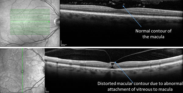
The traction of the vitreous on the macula in Vitreomacular traction syndrome results in a distortion in the normal position of the macula. The normal position of the macula is very important for clear, undistorted vision.
Typically, symptoms of Vitreomacular Traction Syndrome include decreased sharpness of vision, photopsia (flashes of light), micropsia (objects appear to be smaller than they actually are), metamorphopsia (distortion of vision where straight lines appear wavy or disappear). These symptoms can appear slowly or intermittently over time, perhaps with more distortion of vision than loss of sharpness. Some patients might experience some of these symptoms without losing visual acuity.
For this reason, it is important that you contact your retina specialist any time you experience changes to your vision, no matter how mild the changes may seem. Your ophthalmologist or retina specialist will perform a detailed eye examination and will use a diagnostic imaging technique called Optical Coherence Tomography (OCT) to take a scan of the back of your eye. This scan will reveal whether VMT is present and whether it has caused any further pathology. If the scan reveals VMT without any other pathology, your retina specialist will likely decide just to monitor the condition, but this will vary on a case by case basis. Your retina specialist may decide to treat your VMT syndrome if it is causing retinal issues such as a macular hole for example. Again, this will differ from patient to patient and your case and prognosis will be determined by your retina specialist.
If you experience any changes to your vision, do not hesitate to schedule an appointment with a retina specialist. The sooner your condition is evaluated, the better the potential outcome for your vision. Our physicians are taking on new patients, call for a consult today!
In the interest of maintaining further transparency and providing a wide breadth of information to our patients and providers, this blog will serve as an educational and informative resource on interesting happenings within Retina Consultants of Boston and in the greater field of Ophthalmology.
Here at Retina Consultants of Boston, Dr. John J. Weiter and Dr. Namrata Nandakumar are on the forefront of diagnostic techniques, treatment and micro-surgical techniques for macular degeneration, diabetic retinopathy, retinal detachments, macular holes, and a number of other issues affecting the vitreous and retina. Check back here frequently for news and updates on our practice and all things retina!