Make an Appointment (978)-854-5090
By admin | Apr 30, 2018
Blurry vision can be caused by a host of things. Some are relatively benign such as dry eyes or cataracts. Others, however, can be due to conditions that can cause irreversible decline in vision if not addressed immediately. One such condition is called a retinal detachment.
The retina is a light sensitive tissue which embryologically has neural tissue origin. It converts light signals into electrical signals that are then sent to the brain. This means, just like the brain, any damage will cause irreversible loss in vision. There are many different types of retinal detachments. None of them should be taken lightly.
There are different types of retinal detachments:
Rhegmatogenous detachment is caused by a tear or beak in the retina which causes fluid to build up underneath the retina and detach the retina from the pigment cell layer of the retina called the retinal pigment epithelium (RPE). These types of retinal detachments are the most common. It occurs most commonly when the vitreous gel, located in the middle of the eye starts to cross link and separate. This is a normal aging change. Usually in over 90% of cases, this separation is uneventful. However, in some cases, there is a sharp pull on the retina as the vitreous is pulling away, and this is what leads to a retinal tear. If fluid gets under the tear, it leads to a retinal detachment. Light flashes and floaters may signal the onset of the vitreous gel pulling away. It is important to be seen right away if you experience these symptoms because it is not possible to rule out a tear or retinal detachment without a good clinical examination. Caught early, rhegmatogenous retinal detachment can sometimes be repaired in the office with laser or freezing treatments. Even if it needs to be repaired surgically in the operating room, timely intervention is key to saving sight. The sooner the retina is repaired, the better the chance of the tissue trying to heal properly, and the better the chance of preserving your sight.

Tractional detachment is caused when scar tissue is present on the retinas surface. The scar tissue causes contractions on the surface or within the retina. These contractions can cause the retina to separate from its usual position. Some of the most common causes of tractional detachments include advanced diabetes or trauma. Just like with rhegmatogenous detachments, timely intervention is key to trying to preserve vision. The longer the retina stays detached, the less chance there is for the wiring of the retina to recover, and the less the chance of preserving sight.

Exudative detachment occurs when there is no break in the retinal tissue, but fluid gets under the retina. This is commonly due to systemic diseases, inflammation, and leaking blood vessels. Because the retina is not in its proper position, vision becomes blurry. If the fluid is present chronically, it will cause the retina to slowly deteriorate. Therefore, it is important to understand what is causing the exudative detachment and try to address the source problems.

Retinal detachments can occur at any age, but those with a family history of retinal detachments, suffering an injury to the eye, or the presence of retinal diseases such as diabetic retinopathy are more at risk for developing a retinal detachment.
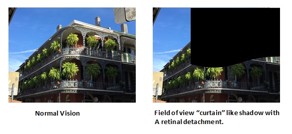
If you suddenly experience flashes of light, floaters or a curtain like shadow in your field of vision contact your ophthalmologist right away. Treatment for a retinal requires timely intervention. Your ophthalmologist will perform a dilated eye exam to better asses the degree of detachment and treatment options. Here at Retina Consultants of Boston, our Board-Certified Ophthalmologist work closely with patients on a case by case basis. We are skilled in managing all types of retinal detachments and use advanced surgical techniques to achieve good surgical outcomes. Our goal is to provide optimized care for our patients and develop a successful treatment plan to best preserve your vision. We are currently accepting new patients, please call and schedule an appointment today.
In the interest of maintaining further transparency and providing a wide breadth of information to our patients and providers, this blog will serve as an educational and informative resource on interesting happenings within Retina Consultants of Boston and in the greater field of Ophthalmology.
Here at Retina Consultants of Boston, Dr. John J. Weiter and Dr. Namrata Nandakumar are on the forefront of diagnostic techniques, treatment and micro-surgical techniques for macular degeneration, diabetic retinopathy, retinal detachments, macular holes, and a number of other issues affecting the vitreous and retina. Check back here frequently for news and updates on our practice and all things retina!